

خرید و دانلود نسخه کامل کتاب Imaging Anatomy: Text and Atlas Volume 3: Bones, Joints, Muscles, Vessels, and Nerves (Atlas of Imaging Anatomy) 1st Edition
قیمت اصلی 177,000 تومان بود.79,000 تومانقیمت فعلی 79,000 تومان است.

خرید و دانلود نسخه کامل کتاب Imaging of Orthopedic Sports Injuries (Medical Radiology) Second Edition 2021
قیمت اصلی 177,000 تومان بود.79,000 تومانقیمت فعلی 79,000 تومان است.
📥 ترافیک اینترنت مصرفی جهت دانلود فایلها در الی فایل، بهصورت نیمبها می باشد.
-55%
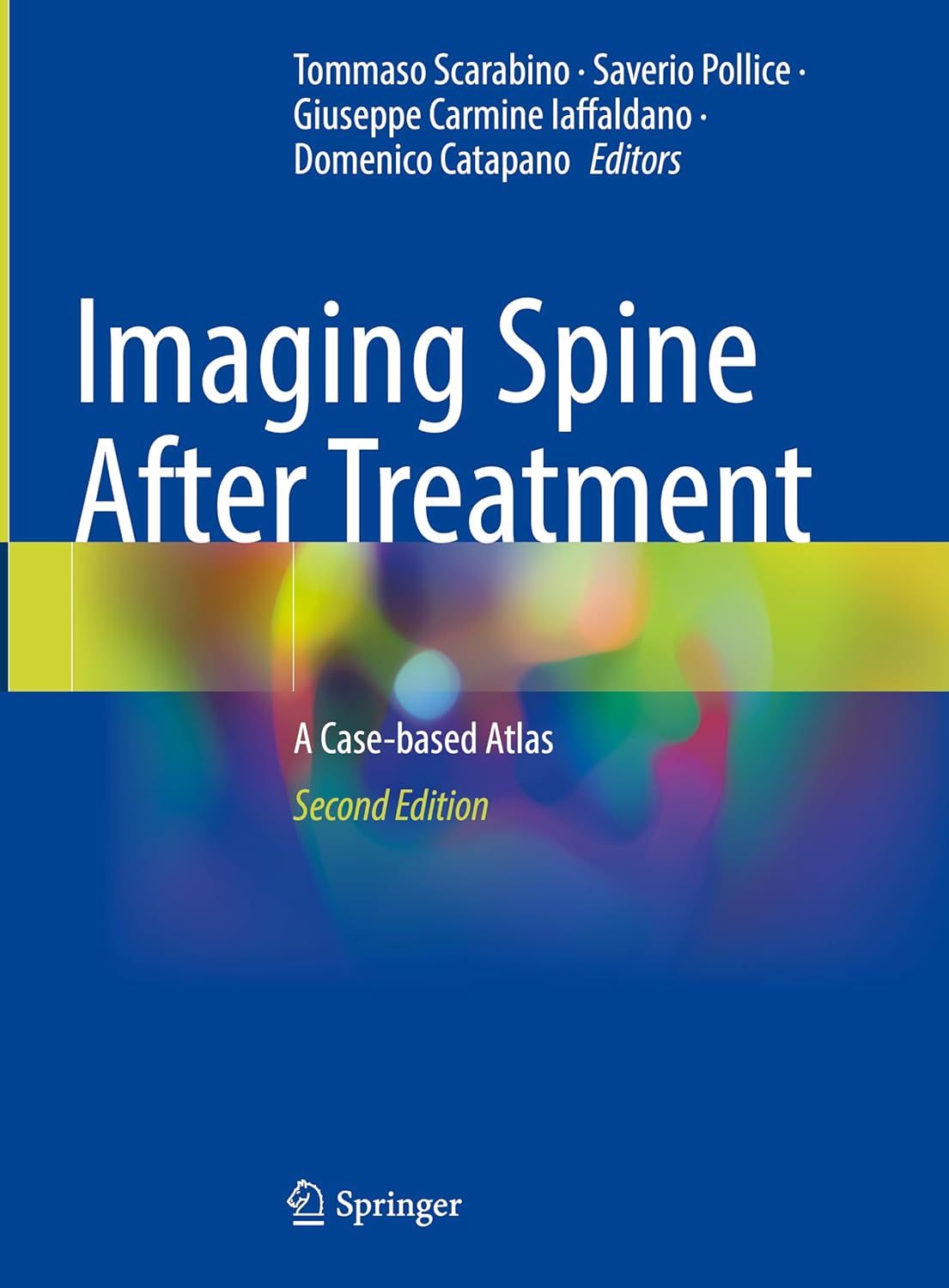
خرید و دانلود نسخه کامل کتاب Imaging Spine After Treatment: A Case-based Atlas Second Edition 2024
قیمت اصلی 177,000 تومان بود.79,000 تومانقیمت فعلی 79,000 تومان است.
تعداد فروش: 67
نویسندگان : Tommaso Scarabino (Editor), Saverio Pollice (Editor), Giuseppe Carmine Iaffaldano (Editor), Domenico Catapano
فرمت فایل :PDF اصلی
تعداد صفحات : 469 صفحه نسخه کامل
زبان انگلیسی
2
آیتم آخرین فروخته شده 55 دقیقه
3
افرادی که اکنون این محصول را تماشا می کنند!
توضیحات
آنتونی رابینز میگه : من در 40 سالگی به جایی رسیدم که برای رسیدن بهش 82 سال زمان لازمه و این رو مدیون کتاب خواندن زیاد هستم.
- زبان فایل : انگلیسی
- نوع فایل : PDF اصلی
توضیحاتی در مورد کتاب
آناتومی تصویربرداری: متن و اطلس جلد 3: استخوانها، مفاصل، عضلات، رگها و اعصاب (اطلس آناتومی تصویربرداری) ویرایش اول توسط فرهود صارمی (نویسنده)، داکش پاتل (نویسنده)، دامیان سانچز-کوئینتانا (نویسنده)، هیرو کیوسو (نویسنده)، منگ لا (نویسنده)، آر. شین تابز (نویسنده)
نظرات (8)
طلا نادری –
الی فایل همیشه در ارائه محتوای باکیفیت پیشرو بوده.
محمدرضا کرمی –
سایت الی فایل محیطی امن و سریع برای خرید فایلهای آموزشی هست.
بهروز محمدی –
پشتیبانی شما در الی فایل همیشه پاسخگو بوده. متشکرم.
پارسا زاهدی –
خیلی وقت بود دنبال این منبع بودم. ممنون از الی فایل که موجودش کرد.
پارسا شاکری –
یک خرید عالی و بیدردسر! حتماً دوباره به الی فایل سر میزنم.
شکیلا صمدی –
محصولی که خریدم بسیار عالی و کاربردی بود. سپاس از شما.
آرمین آذری –
محصولی که خریدم بسیار عالی و کاربردی بود. سپاس از شما.
محمدرضا نادری –
خرید آسان، دانلود سریع، کیفیت عالی. الی فایل کارش درسته.