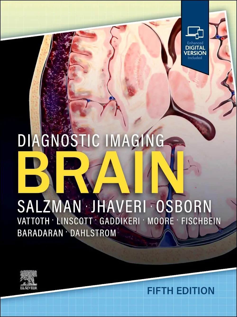
Covering the entire spectrum of this fast-changing field, Diagnostic Imaging: Brain, fifth edition, is an invaluable resource for neuroradiologists, general radiologists, and trainees―anyone who requires an easily accessible, highly visual reference on today’s brain imaging. Drs. Miral Jhaveri, Karen L. Salzman, and a diverse group of neuroimaging experts provide updated information on more than 300 brain and CNS conditions to help you make informed decisions at the point of care. The text is lavishly illustrated, delineated, and referenced, making it a useful learning tool as well as a handy reference for daily practice.
- Provides authoritative, comprehensive guidance on both pathology-based and anatomy-based diagnoses to help you diagnose the full range of brain and CNS conditions
- Contains numerous new diagnoses and new chapters on such topics as inflammatory and demyelinating diseases, metabolic conditions, and CNS tumors
- Reflects significantly revised brain tumor categories based on the WHO CNS Classification and the Consortium to Inform Molecular and Practical Approaches to CNS Tumor Taxonomy, including major changes that advance the role of molecular diagnostics in CNS tumor classification
- Discusses key topics such as advances in treatment of Alzheimer’s disease with the approval of monoclonal antibody (MAB) immunotherapy, CNS manifestations of SARS-CoV-2, and recent advances in vessel wall imaging
- Features nearly 3,200+ high-quality print images and videos (with an additional 4,700+ images in the complimentary eBook), including radiology images, full-color medical illustrations, clinical images, gross pathology photographs, and histology images
- Increases your ability to communicate imaging findings effectively with other neuroscientists, including oncologists and neurosurgeons
- Uses succinct bulleted text and highly templated chapters for quick comprehension of essential information at the point of care
- Includes an eBook that allows you access to everything in the print version as well as new text, videos, and thousands of additional images and references, with the ability to search, customize your content, make notes and highlights, and have content read aloud
توضیحات فارسی در مورد کتاب (ترجمه ماشینی)
«تصویربرداری تشخیصی: مغز، ویرایش پنجم»، یک منبع ارزشمند برای رادیولوژیستهای عصبی، رادیولوژیستهای عمومی و دانشجویان است—هر کسی که به مرجعی بصری و بهراحتی در دسترس برای تصویربرداری مغز در زمان حاضر نیاز دارد. دکترهای میرال جاوهری، کارن ال. سالزمن و گروهی متنوع از متخصصان تصویربرداری عصبی، اطلاعات بهروزرسانی شدهای را در مورد بیش از ۳۰۰ وضعیت مغزی و سیستم عصبی مرکزی ارائه میکنند تا به شما در اتخاذ تصمیمات آگاهانه در محل مراقبت کمک کنند. این متن با تصاویر، طرحها و ارجاعات غنی، نه تنها یک ابزار یادگیری مفید است، بلکه مرجعی مناسب برای عمل روزمره نیز میباشد.
راهنمایی جامع و معتبری را در مورد تشخیصهای مبتنی بر پاتولوژی و آناتومی ارائه میکند تا بتوانید طیف کامل وضعیتهای مغزی و سیستم عصبی مرکزی را تشخیص دهید.
حاوی تشخیصهای جدید و فصلهای جدیدی در مورد موضوعاتی مانند بیماریهای التهابی و دمییلینکننده، شرایط متابولیک و تومورهای سیستم عصبی مرکزی است.
بر اساس طبقهبندی سیستم عصبی مرکزی WHO و کنسرسیوم اطلاعرسانی مولکولی و رویکردهای عملی برای طبقهبندی تومورهای سیستم عصبی مرکزی، دستهبندیهای تومور مغز را بهطور قابل توجهی بازنگری میکند و تغییرات عمدهای را در نقش تشخیص مولکولی در طبقهبندی تومورهای سیستم عصبی مرکزی به جلو میبرد.
موضوعات کلیدی مانند پیشرفتها در درمان بیماری آلزایمر با تایید ایمونوتراپی آنتیبادی مونوکلونال، علائم سیستم عصبی مرکزی SARS-CoV-2 و پیشرفتهای اخیر در تصویربرداری دیواره عروق را مورد بحث قرار میدهد.
حدود ۳۲۰۰ تصویر و ویدیوی با کیفیت بالا (با بیش از ۴۷۰۰ تصویر اضافی در کتاب الکترونیکی همراه) شامل تصاویر رادیولوژی، طرحهای رنگی پزشکی، تصاویر بالینی، عکسهای پاتولوژی بزرگنمایی شده و تصاویر هیستولوژی ارائه میکند.
توانایی شما را در ارتباط موثر نتایج تصویربرداری با سایر دانشمندان اعصاب، از جمله آنکولوژیستها و جراحان مغز و اعصاب، افزایش میدهد.
از متن خلاصهشده با استفاده از لیستهای نقطهای و فصلهای قالببندیشده برای درک سریع اطلاعات ضروری در محل مراقبت استفاده میکند.
یک کتاب الکترونیکی شامل تمام محتوا را در اختیار شما قرار میدهد.


نقد و بررسیها
هنوز بررسیای ثبت نشده است.