

خرید و دانلود نسخه کامل کتاب Tumors and Tumor-Like Lesions of Bone: For Surgical Pathologists, Orthopedic Surgeons and Radiologists 1st ed. 2015 Edition
قیمت اصلی 177,000 تومان بود.79,000 تومانقیمت فعلی 79,000 تومان است.

خرید و دانلود نسخه کامل کتاب Imaging Spine After Treatment: A Case-based Atlas Second Edition 2024
قیمت اصلی 177,000 تومان بود.79,000 تومانقیمت فعلی 79,000 تومان است.
📥 ترافیک اینترنت مصرفی جهت دانلود فایلها در الی فایل، بهصورت نیمبها می باشد.
-55%
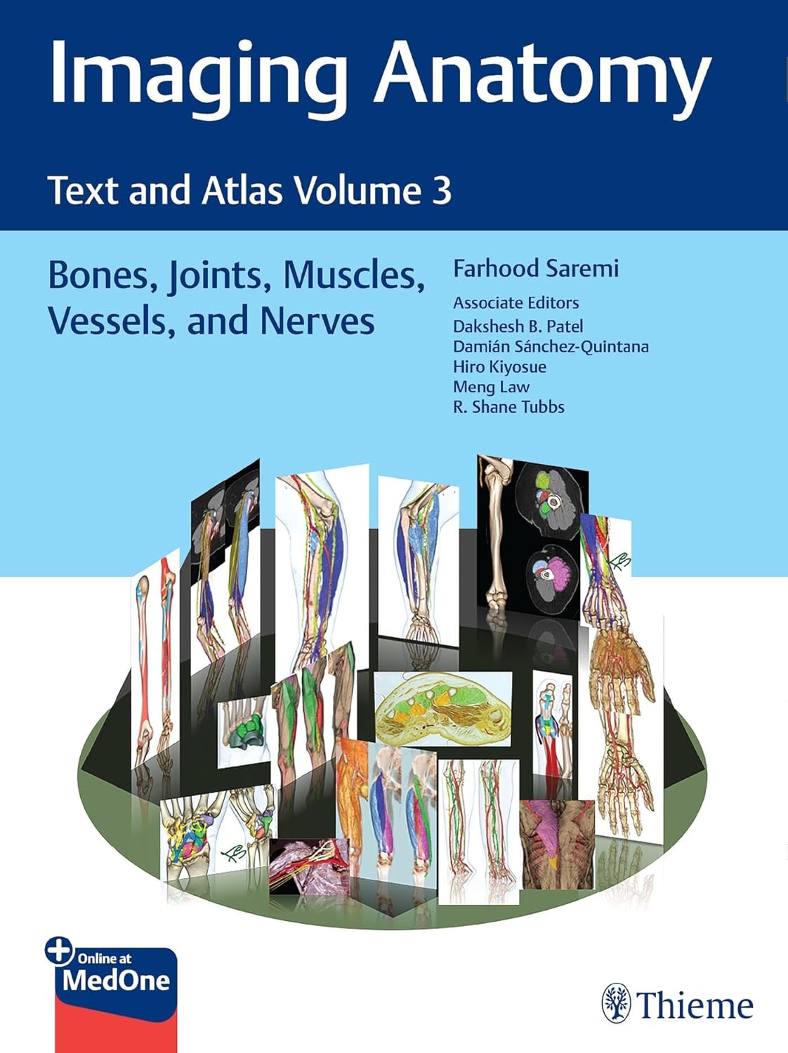
خرید و دانلود نسخه کامل کتاب Imaging Anatomy: Text and Atlas Volume 3: Bones, Joints, Muscles, Vessels, and Nerves (Atlas of Imaging Anatomy) 1st Edition
قیمت اصلی 177,000 تومان بود.79,000 تومانقیمت فعلی 79,000 تومان است.
تعداد فروش: 67
نویسندگان : Farhood Saremi (Author), Dakshesh Patel (Author), Damian Sanchez-Quintana (Author), Hiro Kiyosue (Author), Meng Law (Author), R. Shane Tubbs
فرمت فایل :PDF اصلی
تعداد صفحات : 942 صفحه نسخه کامل
زبان انگلیسی
2
آیتم آخرین فروخته شده 55 دقیقه
5
افرادی که اکنون این محصول را تماشا می کنند!
توضیحات
آنتونی رابینز میگه : من در 40 سالگی به جایی رسیدم که برای رسیدن بهش 82 سال زمان لازمه و این رو مدیون کتاب خواندن زیاد هستم.
- زبان فایل : انگلیسی
- نوع فایل : PDF اصلی
توضیحاتی در مورد کتاب
آناتومی تصویربرداری: متن و اطلس جلد 3: استخوانها، مفاصل، عضلات، رگها و اعصاب (اطلس آناتومی تصویربرداری) ویرایش اول توسط فرهود صارمی (نویسنده)، داکش پاتل (نویسنده)، دامیان سانچز-کوئینتانا (نویسنده)، هیرو کیوسو (نویسنده)، منگ لا (نویسنده)، آر. شین تابز (نویسنده)
نظرات (7)
کوروش اکبری –
واقعاً از خرید این مجموعه راضی هستم. سپاس از الی فایل.
کیانا صمدی –
بسیار عالی و دقیق. ممنون از پشتیبانی سریع و حرفهای الی فایل.
فرزانه کاظمی –
خیلی کاربردی بود، دقیقاً همون چیزی بود که برای پروژهام نیاز داشتم.
شکیلا صابری –
محصولی که خریدم بسیار عالی و کاربردی بود. سپاس از شما.
شکیلا جلالی –
سایت الی فایل از نظر کاربری فوقالعاده است. خرید کتاب از اینجا عالیه.
کتایون فلاح –
کیفیت و دقت کار شما ستودنی است. ممنون الی فایل.
پارسا قاسمی –
خرید آسان، دانلود سریع، کیفیت عالی. الی فایل کارش درسته.